Canna~Fangled Abstracts

Plant flavonoids in cancer chemoprevention: Role in genome stability

By November 14, 2016No Comments
 DOI: 10.1016/j.jnutbio.2016.11.007
Journal of Nutritional Biochemistry
elsevier logo cropped
Available online at: www.sciencedirect.com
ScienceDirect
Journal of Nutritional Biochemistry 45 (2017) 1 14
 

REVIEWS: CURRENT TOPICS

Plant flavonoids in cancer chemoprevention: role in genome stability

Vazhappilly Cijo George (a), Graham Dellaire (b), H.P. Vasantha Rupasinghe (a,b,⁎)
(a)Department of Plant, Food, and Environmental Sciences, Faculty of Agriculture, Dalhousie University, Truro, Nova Scotia, Canada
(b)Department of Pathology, Faculty of Medicine, Dalhousie University, Halifax, Nova Scotia, Canada
Received 3 July 2016; received in revised form 27 August 2016; accepted 14 November 2016

Abstract
Carcinogenesis is a multistage process that involves a series of events comprising of genetic and epigenetic changes leading to the initiation, promotion, and progression of cancer. Chemoprevention is referred to as the use of nontoxic natural compounds, synthetic chemicals or their combinations to intervene in multistage carcinogenesis. Chemoprevention through diet modification, i.e. increased consumption of plant-based food, has emerged as a most promising and potentially cost-effective approach to reducing the risk of cancer. Flavonoids are naturally occurring polyphenols that are ubiquitous in plant-based food such as fruits, vegetables, teas as well as in most medicinal plants. Over 10,000 flavonoids have been characterized over the last few decades. Flavonoids comprise of several sub-classes including flavonols, flavan-3-ols, anthocyanins, flavanones, flavones, isoflavones and proanthocyanidins. This review describes the most efficacious plant flavonoids, including luteolin, epigallocatechin gallate (EGCG), quercetin, apigenin, and chrysin, their hormetic effects, and the molecular basis of how these flavonoids contribute to the chemoprevention with a focus on protection against DNA damage caused by various carcinogenic factors. The present knowledge on the role of flavonoids in chemoprevention can be used in developing effective dietary strategies and natural health products targeted for cancer chemoprevention.
© 2017 Elsevier Inc. All rights reserved.
Keywords: Cancer; Flavonoids; Chemoprevention; Dietary antioxidants; DNA damage signaling and repair; Polyphenols; DNA damage detection; Genome stability

1. Introduction

elsevier-proper-thumbThe term cancer can be described as a set of complex processes involving impaired cells death, unlimited cell proliferation and temporalspatial changes in cell physiology that often leads to malignant tumor formation resulting in invasion of distant tissues to form metastasis [1]. Multistage carcinogenesis is a widely accepted
hypothesis in the development of cancers and is operationally divided into three stages, namely, initiation, promotion and progression [2,3]. Carcinogenesis may result from extensive DNA damage, often caused by exposure to a variety of exogenous and endogenous agents including ultraviolet radiation (UVR), ionizing radiations (IRs), mutagenic chemicals, environmental agents, therapeutic agents or diagnostic imaging. DNA damage as a term encapsulates both frank single and double-stranded DNA breaks, as well as stable modications to nitrogen bases in DNA or its sugar-phosphate backbone, caused by external (e.g., IR) or internal sources [e.g., reactive oxygen species (ROS) generated during oxidative metabolism], which impact the cell by disrupting gene function and/or impairing transcription, DNA replication and cell proliferation [4]. Maintaining genomic integrity is therefore crucial for the organism since it is a key feature in the maintenance of cell function and inappropriate DNA repair is associated with both the initiation and progression of cancer [5]. Failure in proper DNA protection and DNA repair mechanisms, decrease in cellular defenses, malfunctions in cell cycle checkpoints and aberrant inammatory signaling can contribute to poor genomic stability and provide an Achilles heelexploited by many cancer therapeutics [6]. As such, the differences in the DNA damage response between normal and cancer cells often underlie the utility of DNA damaging agents in cancer treatment [7]. DNA damage occurring during Sphase of cell cycle, when DNA is replicated, was considered as the most lethal DNA damage [8] and, given the uncontrolled proliferation of cancer cells, may explain why DNA-damaging agents can be so effective in targeting cancers.
______
Abbreviations: ATM, ataxia telangiectasia mutated; ATR, ATM-Rad3-related; BRCA, breast cancer proteins; CHK, checkpoint kinases; DDR, DNA damage response; DNA-PK, DNA-protein kinases; DRI, dose reduction index; DSBs, double strand breaks; EGCG, epigallocatechin-3-gallate; H2O2, hydrogen peroxide; HDAC, histone deacetylase; HR, homologous recombination; IF, immunouorescence; IR, ionizing radiation; MDC1, mediators of DNA damage checkpoint 1; mTOR, the mechanistic target of rapamycin; NHEJ, nonhomologous end joining; PI, propidium iodide; PI3K, phosphatidylinositol-3 kinases; RAD51/52, radiation-induced assembly; ROS, reactive oxygen species; SSBs, single-strand breaks; UVR, ultraviolet radiation. ⁎Corresponding author. Tel.: +1 902 893 6623; fax: +1 902 893 1404.
E-mail address: vrupasinghe@dal.ca (H.P.V. Rupasinghe).
http://dx.doi.org/10.1016/j.jnutbio.2016.11.007
0955-2863/© 2017 Elsevier Inc. All rights reserved.
 ____
2. Cellular DNA-damaging agents
Cellular DNA can be damaged by cytotoxic or genotoxic agents with different mechanism of action. Some of the classical groups of DNA damage inducers used in chemotherapy are alkylating agents, platinum drugs, antimetabolites, topoisomerase inhibitors and forms of ionizing radiation [9]. Alkylating agents like the nitrogen mustards, nitrosoureas, aziridine compounds, alkyl sulphonates and triazine compounds are electrophiles that covalently transfer alkyl groups onto the DNA bases, disrupting the DNA helix shape [10]. Cisplatin and its analogs, carboplatin and oxaliplatin, are DNA-intrastrand cross-linking agents, and these platinum-based drugs are often used to treat testicular or ovarian cancers [11]. The uoropyrimidine 5-uorouracil, an antimetabolite having similar structures that are related to nucleotide metabolites, induces DNA damage by either inhibiting biosynthetic processes or being incorporated into nucleic acids such as DNA and RNA [12]. Camptothecin, a plant alkaloid, is a topoisomerase inhibitor widely used to treat colorectal, ovarian and lung cancers that targets DNA-TOP1 (type 1 topoisomerase) cleavage complexes, blocking religation and resulting in the accumulation of transient single-strand breaks (SSBs) [13]. A summary of factors which causes DNA damage is depicted in Fig.1.
3. DNA damage response (DDR)
The DDR is a complex cascade of molecular and cellular events that is necessary to eliminate lethal and tumorigenic mutations caused by different genotoxic stress including carcinogens produced by physical or chemical sources. This signaling mechanism regulates cell proliferation, cell cycle and apoptotic induction [14]. Failure in proper DNA damage response mechanisms may result in improper DNA repair which can drive to tumorigenesis and can affect sensitivity to genotoxic chemotherapy [10]. DNA repair can be initiated by various enzymes that modify the DNA and nuclear damage by activating polymerases, topoisomerases, ligases, kinases, phosphatases and glycosylases [15]. DDR is commonly activated in early neoplastic lesions and likely protects against malignancy [16].
4. Signal transduction mechanisms in DNA damage
DNA double-strand breaks (DSBs) are well regarded as the most lethal lesions among all types of damages and possess the greatest challenge to human beings. DSBs can be caused by UV, radiotherapy, dysfunctional telomeres or genotoxic agents, and the breaks they induce can activate phosphatidylinositol-3 kinases (PI3K) including ataxia telangiectasia mutated (ATM), ATM-Rad3-related (ATR) or DNA-dependent protein kinases (DNA-PK) that serve as the pinnacle step in DNA damage signaling (Fig. 2). DNA-PK is a multicomponent complex consisting of the DNA-PK catalytic subunit (DNA-PKcs) and the Lupus Ku autoantigen protein heterodimer (Ku80 and Ku70) [17]. Although there is some cross talk in downstream targets of ATM and ATR, these kinases are activated by ionizing irradiation or ultraviolet light/hydroxyurea, respectively [18]. While DNA-PK regulates a small group of proteins involved in DNA DSB end joining, it also cooperates with ATR and ATM to phosphorylate proteins involved in the DNA damage checkpoints [19]. Thus, protein phosphorylation plays a crucial role in DNA damage signaling, activating over 700 proteins in response to DNA damage which in turn counteract genotoxic stresses by regulating proteinprotein interactions and other post translational modications [20]. However, the functional signicance of many of these proteins is unclear and remains an area of intense research. DNA is packaged within chromatin, and the reversible acetylation of proteins within chromatin-like histone H3 and H4 can affect DNA repair by a number of mechanisms including altering the association of DNA repair factors with damaged DNA and the modulation of the relaxation (or condensation) state of chromatin that may be important in regulating physical access of the DNA repair machinery to the break within chromatin [7,21]. Acetylation is mediated by histone acetyltransferases (or HATs), and deacetylation is mediated by histone deacetylases (HDACs); the balance of HAT and HDAC activities is often perturbed in cancers. Thus, histone acetylation could serve as a therapeutic target for cancer treatment [22]. For example, HDAC inhibitors have the potential to interfere with DNA repair and chromatin relaxation mechanisms, potentially sensitizing cancer cells to DNA damage [23]. Another critical posttranslational modication of chromatin is phosphorylation, and one of the earliest events during DNA DSB repair is the phosphorylation of Ser139 on the specialized histone H2AX called, which is then referred to as γ-H2AX. H2AX is one of the heteromorphous variants of family of at least eight protein species of the nucleosome core histone H2A [24]. Cytologically, γ-H2AX forms punctate structures in the nucleus known as DNA repair foci, which result from the spread of γ-H2AX along chromatin surrounding the DNA break up to 12 megabases of DNA via the action of ATM. These repair foci serve as platforms for the assembly and recruitment of other DNA repair factors, including mediators of DNA damage checkpoint 1 (MDC1) to initiate the DNA damage response [25]Once initiated, the DNA damage signal is amplied by checkpoint kinases 1 and 2 (CHK1 and CHK2) that are activated by phosphorylation by ATM and ATR, and a number of effector proteins, including breast cancer antigens 1 and 2 (BRCA1, BRCA2), p53 and murine double minute [26]. Chk1 and Chk2 can phosphorylate DNA repair proteins like BRCA2 and are involved in cell cycle checkpoint control by phosphorylating proteins such as p53 [27]. BRCA1 and BRCA2 are involved in recruiting the repair protein RAD51 to sites of DNA damage to facilitate DSB repair by homologous recombination (HR) [2830]. The p53 is phosphorylated by Chk1 and Chk2 in response to DNA damage and in turn regulates cell fate decisions including cell cycle arrest and apoptosis by inducing expression of protein such as p21 and B-cell CLL/lymphoma 2 (BCL2), respectively [7,31]. Highlighting the intimate link between DNA repair and cancer, germline or acquired mutations in several DNA repair and signaling factors including BRCA1, BRCA2, ATM, p53 and CHK2 can contribute to the development of cancers affecting multiple organs including breast and ovary, lungs, pancreas and blood (i.e., leukemias) [3235]. Other common syndromes associated with increased cancer susceptibility include Seckel syndrome (mutated ATR), radiosensitive severe combined immunodeciency disease and ligase IV (LIG4) syndrome (mutated LIG4)[36]. After DNA has been repaired, chromatin must be restored to its original state before damage to allow efcient transcription and replication of DNA. Some of the rst steps in chromatin restoration include the dephosphorylation of γ-H2AX by the phosphatases PP4 and PP2A, the proteasomal degradation of MDC1 within repair foci, and deacetylation of H3 or H4 lysines by HDAC [37].Other proteins like the histone chaperones chromatin assembly factor 1 and antisilencing factor 1 also play a crucial role in restoring chromatin structure and cell cycle progression [38]. Failure of these mechanisms may result in epigenetic alterations and thus cause genomic instabilities and its associated diseases [39]. Epigenetic alterations in DNA, such as methylation, and of chromatin, such as modication of histones by acetylation, methylation, and phosphorylation, are increasingly being recognized for their role in health [36,40]. Most of the diseases involving dysregulation of epigenetics are marked with common characteristics such as developmental defects, immunodeciency, neurological degeneration and cancer predisposition [17]. DNA methylation of cytosine (5-methylcytosine) is a very common epigenetic mechanism involved in controlling DNA structure, chromosome stability, the mobility of viral DNA-repeated elements (transposons, retrotransposons), gene imprinting and gene expression [41]< span class="wsc4">. In tumor tissues, tumor suppressor genes are often inactivated epigenetically by methylation of cytosine when compared with normal tissue [42].DNA Damaging Agents
5. Pathways of DNA repair are lesion specific
DNA repair mechanisms are very specic for many types of lesions. Mismatch repair facilitates the replacement of incorrect bases with correct bases. Base excision repair enables to carry small chemical alterations of DNA bases through excision of damaged nitrogen bases [43]. Lesions caused by pyrimidine dimers and intrastrand crosslinks are rectied by removing an oligonucleotide containing the damaged bases through a process called nucleotide excision repair (NER) [44]. SSBs and DSBs are processed by SSB repair and non homologous end joining (NHEJ)/HR mechanism, respectively. Among these mechanisms, NHEJ promotes the potentially inaccurate regulation of DSBs, while HR restores the genomic sequence of the broken DNA ends by employing sister chromatids as a template for repairing DNA [15]. Lupus Ku autoantigen protein p70 (Ku70) binds to DNA DSBs and activates the DNA-PKcs to form DNA-PK holoenzyme which is required for NHEJ pathway of DNA repair [45].
6. Methods to detect DNA damage
There are few traditional methods used to detect DNA damage including neutral sucrose gradients and pulse-eld gel electrophoresis which are laborious, are time consuming and have low sensitivity [46]. Recent research involved much more precise techniques to quantify the DNA damage, and one such common method is single-cell gel electrophoresis (comet assay) that appears to be an attractive method to measure the kinetics of the process including excision followed by resynthesis and ligation of fragments [47,48]. Flavonoids have been studied extensively for their mechanism of action on DNA damage and repair mechanism on various models by using comet assay [49]. As a sensitive measurement of DNA DSBs, phosphorylated histone H2AX protein (γ-H2AX) can be measured by immunouorescence (IF) analysis by using antibodies against γ-H2AX [50]. p53-binding protein 1 (53BP1), MDC1 and PP2A are the other nuclear foci that can detect DNA damage by IF analysis [51]. Double-stranded DNA fragments can also be labeled with terminal deoxynucleotidyl transferase and detected by terminal deoxynucleotidyl transferase dUTP nick end labeling (TUNEL) assay using uorescence microscopy [52].In addition, carcinogen-induced cell death can be measured through multiparameter ow cytometry for apoptosis or necrosis cell death [annexin V/propidium iodide (PI), cell cycle analysis (PI) or cytosolic reactive oxygen species (dichlorouorescein diacetate)] [53]. Various methods used to detect and measure DNA damage and its associated proteins are summarized in Tables 1 and 2.
7. Cancer chemotherapy
Treatment methods for cancers normally involve surgery, radiation therapy, immunotherapy, targeted therapy and chemotherapy with drugs that can kill cancer cells. Chemotherapy for cancer control is based on the presumption that cancer develops through a multistep process. Chemopreventive agents function as either blocking agents, which act immediately before or during the initiation of carcinogenesis by chemical carcinogens, or suppressing agents, which act after initiation during the prolonged stages of promotion and progression [54]. Development of chemopreventive drugs requires specic and/or multiple molecular and cellular targets to act on in order to control proliferation/growth of cancerous cells. Dose selection of chemotherapeutic drug is critical for therapy since some of the anticancer drugs possess systemic toxic effects when used at higher concentrations. In the process of increasing efcacy for chemotherapeutic drugs, increasing dosage may have a reversible pharmacological effect to be cytostatic rather than cytotoxic [55]. Combination of two drugs, often indexed as dose reduction index (DRI), is important in systemic toxicity of drugs. Lower DRI values (N1) may be a good choice as it may protect normal cells and selectively destroys cancer cells [56]. However, a good chemotherapeutic drug at therapeutic dosages ideally targets specic pathways which are aberrant in cancer cells relative to normal cells.
RG Fig 2 Plant Flavonoids

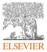
Fig. 2. Cellular mechanisms of flavonoids on DNA damage and repair. Different DNA-damaging agents may form DSBs followed by chromatin modifications in order to initiate damagesignaling proteins and repair mechanisms. These effects can be modulated by flavonoids which inhibit carcinogenesis process by multiple mechanisms including acting as antioxidants,induction of antioxidant defense and phase I/II enzymes, inhibition of DNA damaging proteins, stimulating repair mechanisms in normal cells and restoring genomic integrity in normalcells. Failure of repair mechanisms may result in the uncontrolled cell cycle, somatic mutations (chromosomal abnormalities, telomerase shortening etc.) and malignant tumorformation. Selected examples of flavonoids that are known to regulate carcinogenesis in normal cells: luteolin (1), apigenin (2), EGCG (3), quercetin (4), fisetin (5) and epicatechin (7).Abbreviations: P, phosphorylation; A, acetylation.

Methods to detect DNA damage
7.1. Dietary flavonoids in cancer chemoprevention
Traditional knowledge and scientic reports demonstrate that medicinal plants are rich sources of biologically active compounds that can be used for the treatment of various diseases including some type of cancers. The rst attempt to develop a chemotherapeutic drug began in 1940s with nitrogen mustards and antifolate drugs [57]. Research on avonoids showed major developments in anticancer drug discoveries with potential to destroy cancer cells through apoptotic induction [58,59]. Plants have provided an endless supply of secondary metabolites or phytochemicals that are increasingly exploited against various cancers [60,61]. For example, properties of plant polyphenols including chemical diversity,structural complexity, affordability, lack of substantial toxic effects and inherent biological activities have made them attractive candidates for new therapies [62]. The efcacy of avonoids to act on a target molecule in chemoprevention highly depends on their absorption, metabolism, distribution and availability at the site of action in the body [63,64]. Bioavailability of avonoids is generally low and varies depends on their structure, subclass, molecular weight, glycosylation and esterication. For an instant, intake of dietary avonoid quercetin by humans (250500 mg) may result in b1% dose (unaltered form) that eventually reaches the circulating system with a peak in plasma concentrations in the lower nanomolar range (~20 nM) [65,66]. More often, avonoids get transformed to conjugate metabolites such as Oglucuronides, sulfate esters and O-methyl esters by phase II and hydrolyzing enzymes in the small intestine, liver and colon [67,68]Quercetin glycosides were found to be better absorbed than its counterpart aglycone with observed higher plasma concentrations after being transformed initially in either deglycosylation processes at the intestinal level or carrier-mediated transport [69,70]. These low doses of avonoids may not interfere with cell cycle process in normal cells. However, some compounds like 3-hydroxyavone, luteolin and apigenin when treated with high doses (N50 μM) showed considerable cytotoxicity with increased ROS generation [71]. They may even disturb the cellular integrity by inducing cell death mechanisms in normal cells. It is becoming clear that many external signals including those that stimulate growth, such as growth factors, and those that inhibit growth, such as DNA-damaging agents, control cell proliferations through arresting cell cycle at various phases of cell growth [72]. Current research focuses on modulating effects of these avonoids on cellular signaling pathways involved in DNA damage and repair, which becomes a novel approach in chemoprevention.
7.2. Flavonoids and DNA damage
Studies over a decade from cell cultures, animal and human populations showed the potential of avonoids in human and animal health benets. Since they are abundantly present in our diet, such as fruits, vegetables, teas and wine with strong antioxidative potentials, estrogenic regulatory and antimicrobial activities, they can be exploited against many diseases including cancers [73,74]. They may inhibit several points in cancer progression including invasion, metastasis, angiogenesis, apoptotic mechanisms and cell cycle arrest. Some of the classical examples of avonoids and their mechanism of actions are discussed below (Figs. 3 and 4;Tables 3 and 4).
7.2.1. Apigenin
Apigenin, a avone widely found in many medicinal plants including Lycopodium clavatum L. (club moss) and some vegetables such as Petroselinum crispum L. (parsley) and Apium graveolens L. (celery), has been reported to have benecial effect against UV-B-induced DNA damage and inammatory diseases [75,76]. Recent in vitro and in vivo studies have shown that apigenin possesses antiproliferative properties with induction of cell cycle arrest and apoptosis in cancer models. Apigenin has been reported for DNA protective activity in skin cells and mice against UV-B-induced ROS and DNA damage by removing cyclobutane rings, inhibition of ROS generation and down-regulation of NF-κB[76]. In human diploid broblast and bladder cancer T-24 cells, apigenin showed signicant ROS scavenging potentials along with cell cycle arrest at G1 phase by modulating CDK2 kinases [7779]. Apigenin reduces the risk of
RG Table 2
ovarian cancers as demonstrated by a population study conducted in ovarian cancer patients [80]. Apigenin possess protective effect in endothelial cells from lipopolysaccharide (LPS)-induced inammation. Apigenin reduces ROS production and caspase-3 expression levels and thereby keeps DNA damage in check [81]. In cancer cells, apigenin induces DNA damage/apoptosis by activating ATM and H2AX phosphorylation with down-regulation of cell cycle controlling and DNA repair genes [82]. Apigenin was also reported to modulate DNA damage by inhibiting casein kinase 2, a regulator of cell proliferation, mediator of the DNA damage response and NF-κB activation in malignant glioma cells [83].
7.2.2. Epigallocatechin-3-gallate (EGCG)
An antioxidant potent avan-3-ol, EGCG, protects DNA damage and initiates repair mechanisms in various cancer models. EGCG is the most abundant catechin in green tea and is a traditional anti oxidative free radical scavenger [84]. EGCG was found to be protective in skin cells after irradiation using an X-ray linear accelerator, with overexpression of heme oxygenase-1 (HO-1). EGCG reduces phosphorylation of H2AX foci in these HaCaT keratinocytes [85]. EGCG showed protection against UVR-induced DNA damage in human peripheral blood samples isolated from adult human volunteers before and after drinking 540 ml of green tea [86]. Furthermore, EGCG was also reported to possess radiomodulatory effects on pBR322 plasmid DNA and murine splenocytes against gamma-radiation-induced DNA damage [87].
7.2.3. Luteolin
Luteolin, a avone found in Salvia tomentosa Mill. (Balsamic sage) and many other plants, protects SSBs induced by oxidative stress in PC12 rat pheochromocytoma cells [88]. Luteolin was reported for its apoptotic potentials in human lung squamous carcinoma CH27 cells with higher DNA damage and Sphase cell cycle arrest [89]. Luteolin activates intrinsic apoptotic pathways by inducing DNA damage and p53 in many cancer cells [90,91]. Luteolin induces apoptosis in prostate and breast cancer cells by inhibiting fatty acid synthase, a key lipogenic enzyme overexpressed in many human cancers [59]. The chemopreventive effect and associated mechanisms of luteolin in the JB6 P+ neoplastic mouse cell line and the SKH-1 hairless mouse models were described by Byun et al.[92]. Luteolin has been shown to delay or block the development of cancer cells both in vitro and in vivo, protect DNA from carcinogenic stimulus and induce cell cycle arrest and apoptosis via intrinsic and extrinsic signaling pathways [93]Additionally, luteolin also induces apoptosis in multidrug-resistant cancer cells by ROS generation, DNA damage initiation, activation of ATR/Chk2/p53 signaling, inhibition of NF-kB signaling, activation of p38 and depletion of antiapoptotic proteins [94].
7.2.4. Quercetin
Quercetin glycosides, the avonols abundant in apples, onion and garlic, were reported to induce DNA damage in cancer cells [95]Comparatively, although quercetin has similar structure to kaempferol and luteolin, it was reported to cause more damage to normal mammalian DNA [96]. However, quercetin was also reported to protect DNA damage induced by hydrogen peroxide (H2O2) challenge in Caco-2 human epithelial colorectal adenocarcinoma cells by enhancing DNA repair mechanisms through modulation of DNA repair enzymes [97]. Intraperitoneal administration of quercetin in rats showed protection against radiation-induced DNA damage in kidney and bladder tissues [98]. Its free radical scavenging and antioxidant properties attenuate DNA protection by reducing myeloperoxidase and caspase-3 activities in rats. Quercetin metabolite quercetin-3-O-glucuronide was found to have signicant inhibitory effect on noradrenaline binding to α2-adrenergic receptor by suppressing DNA damage induced by treatment with 4-hydroxyestradiol and noradrenaline in MCF-10A normal human breast cancer cells [99]. Quercetin also reduces γ-H2AX and p53 phosphorylation in HT1080 human brosarcoma cells [100].
7.2.5. Chrysin
Flavonoid, chrysin abundant in Passiora incarnate L. (Maypop) and Oroxylum indicum L. (midnight horrow), was reported to have
Plant Polyphenols
Structure of Plant Falvonoids
protective effect against methylmercury (MeHg)-induced genotoxicity as evidenced by studies conducted using 2-month-old male Wistar rats. The level of glutathione (GSH) in blood was restored in animals administrated with chrysin and inhibits DNA fragmentation induced by methylmercury (MeHg+)[101]. Chrysin reduces the disturbances of redox status, named superoxide dismutase, catalase, glutathione peroxidase and GSH in liver, kidney and brain tissues of rats treated with D-galactose [102]. In another study, chrysin protects free-radical-mediated oxidative stress induced by Nω-nitro-Lariginine methyl ester in male albino rats [103].
7.2.6. Daidzein and genistein
Daidzein and genistein are isoavones found in plants including Glycine max L. (soybean) and Pueraria mirica L. (Kwao Krua) and were reported for their DNA photoprotective effect against UVB-irradiation-induced DNA damage in skin broblasts. They reduce cyclooxygenase-2 and DNA-damage-inducible (Gadd45) genes effectively in treated skin samples. However, the synergistic effect of genistein and daidzein when used in combination provided much more photoprotective effects than single compound treatment [104].
7.2.7. Malvidin
Among dietary anthocyanins, malvidin-3-O-glucoside is one of the major constituents, in particular, in red wine [77]. Malvidin-3-O-glucoside protects endothelial cells against peroxynitrit e-promoted apoptotic death by inhibiting ROS production and apoptotic proteins. Treatment with malvidin-3-O-glucoside also enhances proapoptotic protein expression in endothelial cells [105]. Malvidin was also found to protect DNA damage induced by chemotherapeutic drugs cyclophosphamide, procarbazine and cisplatin in mice. A doseresponse protective effect of malvidin was observed in 30-min pretreated mice against cyclophosphamide-induced chromosomal damage [106].
7.2.8. Citrus flavanones
Hesperetin, a avanone abundant in citrus fruits including oranges and grapefruit, as well as tomatoes and cherries, have anti-inammatory, antioxidant, anticarcinogenic and neuroprotective effects [107]. It protects doxorubicin-induced oxidative stress in rats by reducing DNA fragmentation as evident from the comet and TUNEL assays [108]. Dietary polyphenols like ellagic acid, genistein, emodin and guggulsterone have also been proved to be involved with the regulation of cell cycle and apoptosis in various cancer cells [109]. Another avanone, naringenin, present in orange peel, was found to have the potential to induce DNA damage and apoptosis in HaCaT immortalized keratinocytes and in various other cancer cells [110].
7.3. Flavonoid-rich extracts and DNA damage protection
Plant crude extracts rich with avonoids (quercetin, quercitrin, isoquercitrin and rutin) isolated from the leaves of Scutia buxifolia L. protect from chromosome damage in cultured human lymphocytes by retaining mitotic index and genomic stability against H2O2-induced toxicity [111] (Table 5). An antioxidant-rich plant named Gynostemma pentaphylla (Southern Ginseng) was found to protect human endothelial cells from cholesterol-induced DNA damage by inhibiting excessive ROS production during atherosclerosis. It also inhibits the phosphorylation of H2AX and other PI3K proteins and thereby rendered DNA protection [112]. Flavonoid-rich extracts from rhizomes of Podophyllum hexandrum L. (Himalayan May Apple) were found to protect DNA damage and initiate DNA repair mechanisms in isolated human blood leukocytes. The active components were found to have potentials to inhibit H2AX and P53BP1 phosphorylation during DNA DSBs. It also up-regulates DNA-PKcs and Ku80 to facilitate DNA protection against radiation or assist in DNA repair mechanisms [113]. Withania somnifera L. (Ashwagandha), a commonly used herb in Ayurveda, was recently reported to protect brain DNA damage induced by H2O2 oxidative stress in glioblastoma and neuroblastoma cells through down regulation of γ-H2AX and DSBs proteins such as Rad51, 53BP1 etc.[114]. An ethanolic extract of antioxidant-rich plant Nigella sativa L. was reported to protect radiation-induced DNA damage in Swiss albino mice. Orally fed mice with ethanolic extract of N. sativa showed signicant protection against oxidative injury to spleen and liver as measured by lipid peroxidation and the activity of antioxidant enzymes [115].ExtractsofCrataegus pinnatida L. (Chinese hawberry) pollen were reported to protect from DNA damage in response to H2O2-induced damage in mice lymphocytes. It also exhibits high total phenolic content (17.7±1.0 mg gallic acid equivalent/g), total avonoid content (8.0±1.0 mg rutin/g) and strong free radical scavenging activity which might have rendered for the observed protective effects [116].
7.3.1. Apoptosis induction by flavonoids in cancer cells
Induction of apoptosis in cancer cells by avonoids is associated with their ability to inhibit fatty acid synthase activity [59].Methanolic extracts of Gracilaria tenuistipitata, a genus of red algae, were reported to have anticancer activity in oralsquamous cell carcinoma. The extract induces apoptotic cell death by increasing DNA damage, ROS induction and mitochondrial depolarization. It also increases phosphorylation of γ-H2AX and DSBs, thus enforcing cancer cell to death [117]. Aqueous extract of Fagonia cretica L. (Virgin’s Mantle), used widely as herbal treatment for breast cancer and fever, also induces apoptosis in MCF-7
RG Table 3
and MDA-MB-231 breast cancer cells by inducing γ-H2AX, DNA damage, cell cycle arrest and p53 expression. In the absence of p53, F. cretica extract induces apoptosis by FOXO3a (transcription factor) expression [118]. A schematic representation of different avonoids involved in apoptotic regulation is shown in Fig. 5.
7.3.2. Regulation of mTOR signaling by flavonoids and other polyphenols
The mammalian target of rapamycin (mTOR) is a serine/threonine kinase signaling protein that regulates various intracellular and extracellular mechanisms including cell growth, proliferation, apoptosis and cancer (Fig. 6). Expression levels of mTOR were found to be deregulated in most of the cancer cells, resulting in cell proliferation [119]. Plant polyphenols play a crucial role in inhibiting various kinases including mTOR signaling cascades, which are considered as a therapeutic strategy in cancer prevention. Piceatannol is one such polyphenol present in grapes, berries, peanuts and sugar cane; has natural inhibiting effect on tyrosine and serine/threonine kinases; and was found to regulate mTOR signaling in prostate cancer cells with damaging DNA and apoptosis [119,120]. It also inhibits DU145 and CaP prostate cancer cell proliferations and arrest cell cycle by suppressing CDK activities [121]. Recent research showed that piceatannol rendered protection against H2O2-induced DNA damage but was not involved in DNA repair mechanisms in Burkitt’s lymphoma (Raji) and normal human epidermal keratinocytes (NHEK) [28,122]. Fisetin, a avonol, was also found to inhibit AKT/mTOR signaling in human multiple myeloma and melanoma cells, thereby regulating cell proliferation [123]. Treatment with setin on A549 lung carcinoma cells reduced colony formation with decreased protein expression of PI3K and inhibited Akt or mTOR pathway [124]. Phytochemicals such as boswellic acid, butein and capsaicin have been reported to inhibit PI3K, Akt or mTOR signaling in different cancer cells and are discussed in detail elsewhere [125].
7.4. Flavonoid-rich diet and DNA damage
Increased production of ROS and defective antioxidants/DNA repair mechanisms can damage cellular macromolecules; can lead to changes in chromosome instability, genetic mutation and/or modulation of cell growth; and  may result in cancer. ROS like superoxide radicals, H2O2, hydroxyl radicals, singlet oxygen, peroxyl radical,
RG Table 5
peroxynitrite and nitric oxide can be formed from mitochondria, peroxisomes, inammatory cell activation, exogenous sources including environmental agents, pharmaceuticals and industrial chemicals. Antioxidant-rich diet helps to check the ROS production and to prevent cell and tissue damages [126]. A large number of plant-derived avonoids have been studied as new sources of natural antioxidants which showed the potential to mitigate DNA damage induced by ROS in various cells [63,127]. The best-described property of avonoids is their antioxidant capacity. Since these phenolic compounds can delay, inhibit or prevent the oxidation by scavenging free radicals and reduce oxidative stress. Flavonoid-rich fruit extract derived from Punica granatum L. (pomegranate) was reported to inhibit brain DNA damage against cerebral ischemia/reperfusion injury [128] and blood mononuclear DNA damage in rats [129]. Resveratrol is a polyphenolic compound present in plants including grapes, red wine, nuts, berries and other foods with anti-inammatory, antioxidant and anticancer properties [130]Antioxidant properties of resveratrol were modulated by up-regulating expression of ROS scavengers and phase II enzymes such as superoxide dismutase, catalase, glutathione reductase, glutathione peroxidase, selenophosphate synthase 2, thioredoxin reductase and NAD(P)H:quinone oxidoreductase-1 [131]. During pregnancy, consumption of resveratrol protects mother and fetus from the toxicity induced by environmental pollutants [132]. Berries are among the most widely consumed fruits and are abundant in antioxidant phytochemicals, especially anthocyanins which occur along with other classes of phenolic compounds including ellagitannins, avan-3-ols, procyanidins, avonols and hydroxy-benzoate derivatives. It was found to be effective against cardiovascular disorders, advancing-age-induced oxidative stress, inammatory responses and diverse degenerative diseases [133]. Components of inedible berries can scavenge ROS and reduce oxidative DNA damage, stimulate antioxidant enzymes, inhibit carcinogen-induced DNA adduct formation and enhance DNA repair. Strawberry bioactive compounds are widely known to be powerful antioxidants, and extracts were found to be protective in nature against skin damage induced by H2O2 by improving mitochondrial functionality [134]. Haskap (Lonicera caerulea L.) is a cool climate berry crop which is abundant in polyphenols such as anthocyanins (cyanidin-3-O-glucoside, cyanidin-3,5-diglucoside, peonidin-3-Oglucoside, cyanidin-3-O-rutinoside), hydroxycinnamic acids (chlorogenic acid, 3,5-dicaffeoylquinic acid, neochlorogenic acid), avonols (quercetin-3-O-rutinoside, quercetin-3-O-glucoside) and avan-3-ols (proanthocyanidins, catechins) [135,136]. These diverse components and antioxidative properties of haskap could be used as a source for natural health products [137]. Oral administration in rats with anthocyanin-rich haskap extracts showed radio protective properties [138] and defense against the UVB rays via modulation of antioxidant enzyme activity and reduction of DNA damage in SKH-1 mice [139]. Haskap anthocyanins also protect the oxidation of red blood cell membrane induced by UVB irradiation [140]. Recent research showed that methanolic extract of haskap possesses anti-inammatory activities which are polyphenols-dependent [136]. Kiwifruit, a rich source of vitamin C and other antioxidants, was found to be protective against oxidative DNA damage in in vitro as well as in vivo tests. Consumption of kiwifruit induces resistance to DNA oxidative damage by H2O2 in human lymphocytes [47]. In rats, it induces base-excision repair mechanisms after oral intake for 3 weeks [141]. A study conducted among Korean smokers fed with green vegetable drink (Angelica-keiskei-based juice) supplementation for 8 weeks showed protection in peripheral lymphocytes DNA damage [142], indicating that green vegetable drink exerts cancer-protective effects. Furthermore, a study with six of Taiwan’s indigenous purple-leaved vegetables which are rich in avonoids, anthocyanidins and avonols protects DNA of lymphocytes against H2O2 toxicity [143]. These examples further support the notion that dietary polyphenols play a signicant role in DNA protection from various external and internal stimulants.
8. Hormetic effects of dietary flavonoids and polyphenols
When considering phytochemicals as nutraceuticals in chemoprevention or cancer therapy, it is important to consider that many flavonoids and polyphenols have hormetic effects and in high enough doses are potentially toxic. Hormesis is dened as a biphasic doseresponse phenomenon characterized by low-dose benecial or stimulatory effects and high-dose detrimental or inhibitory effects of a compound on a biological process or phenotype, often depicted as a U-shaped doseresponse curve [144]. The U-shaped, biphasic response arises from the benets of adaptation to a cellular stress at low intensity or levels, and as the stressor increases (e.g., increased levels of a dietary avonoid), the adaptive response no longer compensates for the detrimental effects of the stressor, and the treatment is damaging. To illustrate this concept, we can use the example of the
RG Figure 5

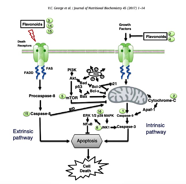
Fig. 5. Schematic illustration of flavonoids in apoptotic pathways in cancer cells. The different modes of action of flavonoids in extrinsic and intrinsic pathways which ultimately lead tocell death. Examples of flavonoid mediators: luteolin (1), apigenin (2), EGCG (3), quercetin (4), fisetin (5), chrysin (6), epicatechin (7), silibinin (14), EGC (15) and myricetin (16).

RG Table 6 a

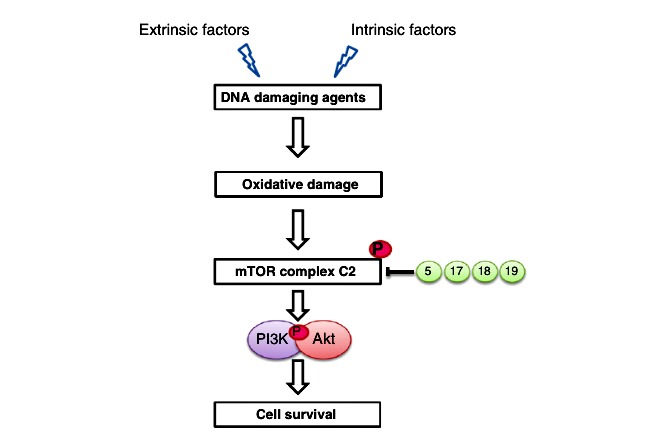
Fig. 6. Graphical representation of the influence of polyphenols in mTOR signaling in cancer cells. Expression levels of mTOR signaling activate continuous cell division and may lead tometastatic conditions. Flavonoids and other phytochemicals inhibit this process by dephosphorylation of mTOR complex formation associated with PI3K and Akt proteins, therebypromoting apoptotic cell death. Examples of polyphenol mediators: fisetin (5), piceatannol (17), caffeic acid (18) and butein (19).

dietary avonoid resveratrol, which has shown biphasic effects in a number of animal models of human disease including gastric lesions, ischemic stroke, osteoporosis and both Alzheimer’s and cardiovascular disease [145]. For example, at low dose, resveratrol can promote cardiac health by promoting the expression of pro survival and antiapoptotic proteins that can improve postischemic ventricular recovery and reduce infarction size following heart attack, whereas the opposite is true for high-dose resveratrol, which promotes cardiomyocyte apoptosis and increases infarct size [146]. With respect to cancer, resveratrol has the striking feature of promoting growth of many cancers of varying histological origin and stage at low dose but exhibiting proapoptotic effects on cancer cells at elevated doses [144]. Resveratrol is more the rule than the exception among dietary flavonoids and polyphenols, and many others including genistein, apigenin, luteolin and kaempferol have all been demonstrated to show hormetic effects by promoting cancer growth at low dose and inhibiting growth at high dose [147]. Similarly, the cancer-preventative effects of quercetin are also dose dependent, being primarily an antioxidant at low doses and a pro-oxidant capable of promoting genomic instability at high doses [148]. Furthermore, the combinatorial activity of multiple dietary avonoids or polyphenols could be strongly additive or synergistic, causing a shift to detrimental or toxic effects on biological processes at relatively low doses not expected to be toxic. These combinatorial hormetic effects should not be taken lightly and may lead to adverse effects in individuals taking health supplements, and are one possible confounding factor in clinical trials where patients enrolled may be concurrently supplementing with over-the-counter nutraceuticals that alter drug clearance, absorption and/or cellular responses. Natural does not equate to safe at all doses and combinations; thus, chemopreventive effects of flavonoids in relation to dose require further investigations.
9. Conclusions and future directions
Human cancers, caused by invariable DNA damage, have posed an increasing global health care concern for over a decade. Various modern treatment methods like targeted therapies have their own limitations including cancer cells developing resistance against anticancer drugs or relapse of cancer after treatment. Hence, there is a need to develop an effective method to treat the uncontrolled cell growth which is often driven by modications of base pairs in DNA. Studies discussed in this review give an insight on how dietary agents, especially avonoids, help to reduce DNA damage and thereby prevent mutations and consequently restore genomic stability. A wealth of scientic evidence is available to demonstrate that avonoids protect from DNA damage induced by various toxic agents. However, there is a lack of knowledge regarding the effects of avonoid-rich diet in modulation of DNA damage prevention/DNA repair mechanisms in healthy cells. Setting an optimum concentration of avonoids is of high importance from a treatment point of view since higher concentrations may also lead to the destruction of normal cells during therapy. Selective nature of some avonoids to protect normal cells and induce cell death mechanisms in cancer cells during chemotherapy or radiotherapy makes them an attractive agent in drug discovery process. However, more studies need to be carried out to congure the mechanism of action of this selective nature of avonoids in order to improve understanding of various epigenetic process which may provide a more rational basis for combining specic dietary compounds and thereby enhancing efcacy in the clinical setting. Furthermore, hormetic effects of dietary avonoids and polyphenols (and their combinations) need to be carefully characterized to prevent unintended side effects, including increasing DNA damage through mechanisms such as ROS production, inhibition or promotion of autophagy required to clear toxic biomolecules and/or down-regulation of important prosurvival pathways to cell stress. In addition, more research should be done with new approaches like modifying chemical structures of avonoids that can increase bioavailability and effectiveness in protection of DNA damage to improve biosafety prole for the dose-responsive effects of avonoids. At the same time, the exploitation of new avonoids which are effective against carcinogens not only contributes to prevent DNA damage but also aids in developing efcient therapeutic regimes. Conflict of interest The authors declare that there is no conict of interest regarding this article.
Acknowledgment
The authors acknowledge the funding provided by the Discovery Grant program of Natural Sciences and Engineering Research Council of Canada (GD and HPVR).
References
[1] Seyfried TN, Shelton LM. Cancer as a metabolic disease. Nutr Metab (Lond) 2010:7.
[2] Trosko JE, Kang KS. Evolution of energy metabolism, stem cells and cancer stem cells: how the Warburg and Barker hypotheses might be linked. Int J Stem Cells 2012;5:3956.
[3] Block KI, Gyllenhaal C, Lowe L, Amedei A, Amin AR, Amin A, et al. Designing a broad-spectrum integrative approach for cancer prevention and treatment. Semin Cancer Biol 2015(Suppl. 35):S276304.
[4] Wajed SA, Laird PW, DeMeester TR. DNA methylation: an alternative pathway to cancer. Ann Surg 2001;234:1020.
[5] Weitzman MD, Lilley CE, Chaurushiya MS. Genomes in conflict: maintaining
genome integrity during virus infection. Annu Rev Microbiol 2010;64:6181.
[6] B DA, H WC, P DS. Destabilization of the cancer genome. Cancer: principles & practice of oncology8 ed. ; 2008.
[7] Rajendran P, Ho E, Williams DE, Dashwood RH. Dietary phytochemicals, HDAC inhibition,and DNA damage/repair defectsin cancer cells.Clin Epigenetics 2011;3:4.
[8] Cheung-Ong K, Giaever G, Nislow C. DNA-damaging agents in cancer
chemotherapy: serendipity and chemical biology. Chem Biol 2013;20:64859.
[9] Woods D, Turchi JJ. Chemotherapy induced DNA damage response: convergence of drugs and pathways. Cancer Biol Ther 2013;14:37989.
[10] Helleday T, Petermann E, Lundin C, Hodgson B, Sharma RA. DNA repair pathways as targets for cancer therapy. Nat Rev Cancer 2008;8:193204.
[11] Kelland L. The resurgence of platinum-based cancer chemotherapy. Nat Rev
Cancer 2007;7:57384.
[12] Longley DB, Harkin DP, Johnston PG. 5-Fluorouracil: mechanisms of action and clinical strategies. Nat Rev Cancer 2003;3:3308.
[13] Pommier Y. Topoisomerase I inhibitors: camptothecins and beyond. Nat Rev
Cancer 2006;6:789802.
[14] Zhou BBS, Elledge SJ. The DNA damage response: putting checkpoints in
perspective. Nature 2000;408:4339.
[15] Ciccia A, Elledge SJ. The DNA damage response: making it safe to play with
knives. Mol Cell 2010;40:179204.
[16] Bartkova J, Horejsi Z, Koed K, Kramer A, Tort F, Zieger K, et al. DNA damage response as a candidate anti-cancer barrier in early human tumorigenesis. Nature 2005;434:86470.
[17] Pandita TK, Richardson C. Chromatin remodeling finds its place in the DNA
double-strand break response. Nucleic Acids Res 2009;37:136377.
[18] StokesMP,RushJ,MacNeillJ,RenJM,SprottK,NardoneJ,etal.ProfilingofUV-induced ATM/ATR signaling pathways. Proc Natl Acad Sci U S A 2007;104:1985560.
[19] Jette N, Lees-Miller SP. The DNA-dependent protein kinase: a multifunctional protein kinase with roles in DNA double strand break repair and mitosis. Prog Biophys Mol Biol 2015;117:194205.
[20] Matsuoka S, Ballif BA, Smogorzewska A, McDonald III ER, Hurov KE, Luo J, et al. ATM and ATR substrate analysis reveals extensive protein networks responsive to DNA damage. Science 2007;316:11606.
[21] Gong FD, Miller KM. Mammalian DNA repair: HATs and HDACs make their mark through histone acetylation. Mutat Res-Fund Mol M 2013;750:2330.
[22] Barneda-Zahonero B, Parra M. Histone deacetylases and cancer. Mol Oncol 2012; 6:57989.
[23] Kachhap SK, Rosmus N, Collis SJ, Kortenhorst MSQ, Wissing MD, Hedayati M, et al. Downregulation of homologous recombination DNA repair genes by HDAC inhibition in prostate cancer is mediated through the E2F1 transcription factor. PLoS One 2010;5.
[24] Bonisch C, Hake SB. Histone H2A variants in nucleosomes and chromatin: more or less stable? Nucleic Acids Res 2012;40:1071941.
[25] Harper JW, Elledge SJ. The DNA damage response: ten years after. Mol Cell 2007; 28:73945. 11V.C. George et al. / Journal of Nutritional Biochemistry 45 (2017) 114
[26] Petrini JH. Cell signaling. A touching response to damage. Science 2007;316: 11389.
[27] Zannini L, Delia D, Buscemi G. CHK2 kinase in the DNA damage response and beyond. J Mol Cell Biol 2014;6:44257.
[28] Bhattacharyya A, Ear US, Koller BH, Weichselbaum RR, Bishop DK. The breast cancer susceptibility gene BRCA1 is required for subnuclear assembly of Rad51 and survival following treatment with the DNA cross-linking agent cisplatin. J Biol Chem 2000;275:23899903.
[29] Dray E, Etchin J, Wiese C, Saro D, Williams GJ, Hammel M, et al. Enhancement of RAD51 recombinase activity by the tumor suppressor PALB2. Nat Struct Mol Biol 2010;17:12559.
[30] Buisson R, Dion-Cote AM, Coulombe Y, Launay H, Cai H, Stasiak AZ, et al.
Cooperation of breast cancer proteins PALB2 and piccolo BRCA2 in stimulating
homologous recombination. Nat Struct Mol Biol 2010;17:124754.
[31] Meek DW. Thep53 response to DNA damage. DNA Repair(Amst) 2004;3:104956.
[32] Nevanlinna H, Bartek J. The CHEK2 gene and inherited breast cancer
susceptibility. Oncogene 2006;25:59129.
[33] Solomon S, Das S, Brand R, Whitcomb DC. Inherited pancreatic cancer
syndromes. Cancer J 2012;18:48591.
[34] McBride KA, Ballinger ML, Killick E, Kirk J, Tattersall MH, Eeles RA, et al. Li-Fraumeni syndrome: cancer risk assessment and clinical management. Nat Rev Clin Oncol 2014;11:26071.
[35] O’Sullivan CC, Moon DH, Kohn EC, Lee JM. Beyond breast and ovarian cancers: PARP inhibitors for BRCA mutation-associated and BRCA-like solid tumors. Front Oncol 2014;4:42.
[36] Langie SA, Koppen G, Desaulniers D, Al-Mulla F, Al-Temaimi R, Amedei A, et al. Causes of genome instability: the effect of low dose chemical exposures in
modern society. Carcinogenesis 2015;36(Suppl. 1):S6188.
[37] Ayoub N, Rajendra E, Su X, Jeyasekharan AD, Mahen R, Venkitaraman AR. The carboxyl terminus of Brca2 links the disassembly of Rad51 complexes to mitotic entry. Curr Biol 2009;19:107585.
[38] Chen CC, Carson JJ, Feser J, Tamburini B, Zabaronick S, Linger J, et al. Acetylated lysine 56 on histone H3 drives chromatin assembly after repair and signals for the completion of repair. Cell 2008;134:23143.
[39] Bishop KS, Ferguson LR. The interaction between epigenetics, nutrition and the development of cancer. Nutrients 2015;7:92247.
[40] Shukla S, Meeran SM, Katiyar SK. Epigenetic regulation by selected dietary
phytochemicals in cancer chemoprevention. Cancer Lett 2014;355:917.
[41] Berman BP, Weisenberger DJ, Aman JF, Hinoue T, Ramjan Z, Liu Y, et al.
Regions of focal DNA hypermethylation and long-range hypomethylation in
colorectal cancer coincide with nuclear lamina-associated domains. Nat Genet 2012;44:406.
[42] Lahtz C, Pfeifer GP. Epigenetic changes of DNA repair genes in cancer. J Mol Cell Biol 2011;3:518.
[43] Jiricny J. The multifaceted mismatch-repair system. Nat Rev Mol Cell Biol 2006;7: 33546.
[44] Hoeijmakers JHJ. DNA damage, aging, and cancer. (vol 361, p 1475, 2009). N Engl J Med 2009;361:1914.
[45] Bennardo N, Cheng A, Huang N, Stark JM. Alternative-NHEJ is a mechanis-
tically distinct pathway of mammalian chromosome break repair. PLoS Genet 2008;4.
[46] SvetlovaMR, Solovjeva LV,Tomilin NV. Application of new methods for detection of DNA damage and repair. Int Rev Cel Mol Biol 2009;277:21751.
[47] Collins AR, Dusinska M, Horvathova E, Munro E, Savio M, Stetina R. Inter-
individual differences in repair of DNA base oxidation, measured in vitro with
the comet assay. Mutagenesis 2001;16:297301.
[48] Collins AR. Investigating oxidative DNA damage and its repair using the comet assay. Mutat Res 2009;681:2432.
[49] Charles C, Chemais M, Stevigny C, Dubois J, Nachergael A, Duez P. Measurement of the influence of flavonoids on DNA repair kinetics using the comet assay. Food Chem 2012;135:297481.
[50] Schmid TE, Zlobinskaya O, Multhoff G. Differences in phosphorylated histone H2AX foci formation and removal of cells exposed to low and high linear energy transfer radiation. Curr Genomics 2012;13:41825.
[51] Mailand N, Bekker-Jensen S, Faustrup H, Melander F, Bartek J, Lukas C, et al. RNF8 ubiquitylates histones at DNA double-strand breaks and promotes assembly of repair proteins. Cell 2007;131:887900.
[52] Hilchie AL,Doucette CD, PintoDM, Patrzykat A, DouglasS, Hoskin DW. Pleurocidin-family cationic antimicrobial peptides are cytolytic for breast carcinoma cells and prevent growth of tumor xenografts. Breast Cancer Res 2011;13.
[53] Chaurio RA, Munoz LE, Maueroder C, Janko C, Harrer T, Furnrohr BG, et al. The progression of cell death affects the rejection of allogeneic tumors in
immune-competent mice – implications for cancer therapy. Front Immunol
2014;5:560.
[54] Gescher A, Pastorino U, Plummer SM, Manson MM. Suppression of tumour
development by substances derived from the diet-mechanisms and clinical
implications. Br J Clin Pharmacol 1998;45:112.
[55] De Angelis C. Side effects related to systemic cancer treatment: are we changing the promethean experience with molecularly targeted therapies? Curr Oncol 2008;15:1989.
[56] Chou TC. Drug combination studies and their synergy quantification using the Chou-Talalay method. Cancer Res 2010;70:4406.
[57] Chabner BA, Roberts Jr TG. Timeline: chemotherapy and the war on cancer. Nat Rev Cancer 2005;5:6572.
[58] Monasterio A, Urdaci MC, Pinchuk IV, Lopez-Moratalla N, Martinez-Irujo JJ. Flavonoids induce apoptosis in human leukemia U937 cells through caspase-
and caspase-calpain-dependent pathways. Nutr Cancer 2004;50:90100.
[59] Brusselmans K, Vrolix R, Verhoeven G, Swinnen JV. Induction of cancer cell
apoptosis by flavonoids is associated with their ability to inhibit fatty acid synthase activity. J Biol Chem 2005;280:563645.
[60] Hussain MS, Fareed S, Ansari S, Rahman MA, Ahmad IZ, Saeed M. Current
approaches toward production of secondary plant metabolites. J Pharm Bioallied
Sci 2012;4:1020.
[61] Fernando F, Rupasinghe HPV. Anticancer properties of phytochemicals present in medicinal plants of North America. Using old solutions to new problems natural drug discovery in the 21st century. Croatia: InTech – Open Access Publisher; 2013.
[62] Ziaullah, Rupasinghe HPV. Application of NMR spectroscopy in plant phenolics associated with human health. Application of NMR spectroscopy in food sciences. Oak Park, IL, USA: Bentham Science; 2015.
[63] Jones QR, Warford J, Rupasinghe HPV, Robertson GS. Target-based selection of flavonoids for neurodegenerative disorders. Trends Pharmacol Sci 2012;33: 60210.
[64] Thilakarathna SH, Rupasinghe HPV. Flavonoid bioavailability and attempts for bioavailability enhancement. Nutrients 2013;5:336787.
[65] Chow HH, Hakim IA. Pharmacokinetic and chemoprevention studies on tea in humans. Pharmacol Res 2011;64:10512.
[66] Marin L, Miguelez EM, Villar CJ, Lombo F. Bioavailability of dietary polyphenols and gut microbiota metabolism: antimicrobial properties. Biomed Res Int 2015; 2015:905215.
[67] Landete JM. Updated knowledge about polyphenols: functions, bioavailability, metabolism, and health. Crit Rev Food Sci Nutr 2012;52:93648.
[68] Kim DH. Gut microbiota-mediated drug-antibiotic interactions. Drug Metab
Dispos 2015;43:15819.
[69] Graefe EU, Wittig J, Mueller S, Riethling AK, Uehleke B, Drewelow B, et al.
Pharmacokinetics and bioavailability of quercetin glycosides in humans. J Clin
Pharmacol 2001;41:4929.
[70] Manach C, Williamson G, Morand C, Scalbert A, Remesy C. Bioavailability and bioefficacy of polyphenols in humans. I. Review of 97 bioavailability studies. Am J Clin Nutr 2005;81:230S42S.
[71] Matsuo M, Sasaki N, Saga K, Kaneko T. Cytotoxicity of flavonoids toward cultured normal human cells. Biol Pharm Bull 2005;28:2539.
[72] Shanmugam MK, Kannaiyan R, Sethi G. Targeting cell signaling and apoptotic pathways by dietary agents: role in the prevention and treatment of cancer. Nutr Cancer 2011;63:16173.
[73] Rupasinghe HPV, Nair S, Robinson R. Chemopreventive properties of fruit
phenolics and their possible mode of actions. In: Studies in natural products
chemistry. Amsterdam: Elsevier Science Publishers; 2014.
[74] Arumuggam N, Bhowmick NA, Rupasinghe HPV. A review: phytochemicals
targeting JAK/STAT signaling and IDO expression in cancer. Phytother Res 2015; 29:80517.
[75] Meyer H, Bolarinwa A, Wolfram G, Linseisen J. Bioavailability of apigenin from apiin-rich parsley in humans. Ann Nutr Metab 2006;50:16772.
[76] Das S, Das J, Paul A, Samadder A, Khuda-Bukhsh AR. Apigenin, a bioactive
flavonoid from Lycopodium clavatum, stimulates nucleotide excision repair
genes to protect skin keratinocytes from ultraviolet B-induced reactive oxygen
species and DNA damage. J Acupunct Meridian Stud 2013;6:25262.
[77] Mazza G. Anthocyanins in grapes and grape products. Crit Rev Food Sci Nutr 1995;35:34171.
[78] Birt DF, Mitchell D, Gold B, Pour P, Pinch HC. Inhibition of ultraviolet light
induced skin carcinogenesis in SKH-1 mice by apigenin, a plant flavonoid. Anticancer Res 1997;17:8591.
[79] Shi MD, Shiao CK, Lee YC, Shih YW. Apigenin, a dietary flavonoid, inhibits
proliferation of human bladder cancer T-24 cells via blocking cel l cycle
progression and inducing apoptosis. Cancer Cell Int 2015;15:33.
[80] Gates MA, Vitonis AF, Tworoger SS, Rosner B, Titus-Ernstoff L, Hankinson SE, et al. Flavonoid intake and ovarian cancer risk in a population-based casecontrol study. Int J Cancer 2009;124:191825.
[81] Duarte S, Arango D, Parihar A, Hamel P, Yasmeen R, Doseff AI. Apigenin protects endothelial c ells from lipopolysacchar ide (LPS)-induced inflamm ation by decreasing caspase-3 activation and modulating mitochondrial function. Int J
Mol Sci 2013;14:1766479.
[82] Arango D, Parihar A, Villamena FA, Wang L, Freitas MA, Grotewold E, et al.
Apigenin induces DNA damage through the PKCdelta-dependent activation of
ATM and H2AX causing down-regulation of genes involved in cell cycle control
and DNA repair. Biochem Pharmacol 2012;84:157180.
[83] Kroonen J, Artesi M, Capraro V, Nguyen-Khac MT, Willems M, Chakravarti A, et al. Casein kinase 2 inhibition modulates the DNA damage response but fails to radiosensitize malignant glioma cells. Int J Oncol 2012;41:77682.
[84] Yin ST, Tang ML, Su L, Chen L, Hu P, Wang HL, et al. Effects of epigallocatechin-3-gallate on lead-induced oxidative damage. Toxicology 2008;249:4554.
[85] Zhu W, Xu J, Ge YY, Cao H, Ge X, Luo JD, et al. Epigallocatechin-3-gallate (EGCG) protects skin cells from ionizing radiation via heme oxygenase-1 (HO-1)
overexpression. J Radiat Res (Tokyo) 2014;55:105665.
[86] Morley N, Clifford T, Salter L, Campbell S, Gould D, Curnow A. The green tea polyphenol ()-epigallocatechin gallate and green tea can protect human
cellular DNA from ultraviolet and visible radiation-induced damage. Photo-
dermatol Photoimmunol Photomed 2005;21:1522.
12 V.C. George et al. / Journal of Nutritional Biochemistry 45 (2017) 114
[87] Richi B, Kale RK, Tiku AB. Radio-modulatory effects of green tea catechin EGCG on pBR322 plasmid DNA and murine splenocytes against gamma-radiation induced damage. Mutat Res 2012;747:6270.
[88] Silva JP, Gomes AC, Coutinho OP. Oxidative DNA damage protection and repair by polyphenolic compounds in PC12 cells. Eur J Pharmacol 2008;601:5060.
[89] Leung HW, Wu CH, Lin CH, Lee HZ. Luteolin induced DNA damage leading to human lung squamous carcinoma CH27 cell apoptosis. Eur J Pharmacol 2005;
508:7783.
[90] Shi R, Huang Q, Zhu X, Ong YB, Zhao B, Lu J, et al. Luteolin sensitizes the
anticancer effect of cisplatin via c-Jun NH2-terminal kinase-mediated p53
phosphorylation and stabilization. Mol Cancer Ther 2007;6:133847.
[91] George VC, Naveen Kumar DR, Suresh PK, Kumar S, Kumar RA. Comparative studies to evaluate relative in vitro potency of luteolin in inducing cell cycle arrest and apoptosis in HaCaT and A375 cells. Asian Pac J Cancer Prev 2013;14: 6317.
[92] Byun S, Lee KW, Jung SK, Lee EJ, Hwang MK, Lim SH, et al. Luteolin inhibits protein kinase C(epsilon) and c-Src activities and UVB-induced skin cancer. Cancer Res 2010;70:241523.
[93] Seelinger G, Merfort I, Wolfle U, Schempp CM. Anti-carcinogenic effects of the flavonoid luteolin. Molecules 2008;13:262851.
[94] Rao PS, Satelli A, Moridani M, Jenkins M, Rao US. Luteolin induces apoptosis in multidrug resistant cancer cells without affecting the drug transporter function: involvement of cell line-specific apoptotic mechanisms. Int J Cancer 2012;130: 270314.
[95] Sudan S, Rupasinghe HPV. Quercetin-3-O-glucoside induces human DNA
topoisomerase II inhibition, cell cycle arrest and apoptosis in hepatocellular
carcinoma cells. Anticancer Res 2014;34:16919.
[96] Yamashita N, Tanemura H, Kawanishi S. Mechanism of oxidative DNA damage induced by quercetin in the presence of Cu(II). Mutat Res 1999;425:10715.
[97] Min K, Ebeler SE. Quercetin inhibits hydrogen peroxide-induced DNA damage and enhances DNA repair in Caco-2 cells. Food Chem Toxicol 2009;47:271622.
[98] Ozyurt H, Cevik O, Ozgen Z, Ozden AS, Cadirci S, Elmas MA, et al. Quercetin protects radiation-induced DNA damage and apoptosis in kidney and bladder tissues of rats. Free Radic Res 2014;48:124755.
[99] Yamazaki S, Sakakibara H, Takemura H, Yasuda M, Shimoi K. Quercetin-3-Oglucronide inhibits noradrenaline binding to alpha(2)-adrenergic receptor, thus suppressing DNA damage induced by treatment with 4-hydroxyestradiol and noradrenaline in MCF-10A cells. J Steroid Biochem Mol Biol 2014;143:1229.
[100] Sun B, Ross SM, Trask OJ, Carmichael PL, Dent M, White A, et al. Assessing dose-dependent differences in DNA-damage, p53 response and genotoxicity for quercetin and curcumin. Toxicol In Vitro 2013;27:187787.
[101] Manzolli ES, Serpeloni JM, Grotto D, Bastos JK, Antunes LM, Barbosa Junior F, et al. Protective effects of the flavonoid chrysin against methylmercury-induced genotoxicity and alterations of antioxidant status, in vivo. Oxid Med Cell Longev 2015;2015:602360.
[102] Anand KV, Mohamed Jaabir MS, Thomas PA, Geraldine P. Protective role of chrysin against oxidative stress in D-galactose-induced aging in an experimental rat model. Geriatr Gerontol Int 2012;12:74150.
[103] T M, R V. Effects of chrysin on free radicals and enzymatic antioxidants in Nωnitro-l-arginine methyl ester: induced hypertensive rats. Int J Nutr Pharmacol Neurol Dis 2014;4:1127.
[104] Iovine B, Iannella ML, Gasparri F, Monfrecola G, Bevilacqua MA. Synergic effect of genistein and daidzein on UVB-induced DNA damage: an effective photoprotective combination. J Biomed Biotechnol 2011;2011:692846.
[105] Paixao J, Dinis TC, Almeida LM. Protective role of malvidin-3-glucoside on
peroxynitrite-induced damage in endothelial cells by counteracting reactive
species formation and apoptotic mitochondrial pathway. Oxid Med Cell Longev
2012;2012:428538.
[106] Khandelwal N, Abraham SK. Protective effects of common anthocyanidins
against genotoxic damage induced by chemotherapeutic drugs in mice. Planta
Med 2014;80:127883.
[107] Kara S, Gencer B, Karaca T, Tufan HA, Arikan S, Ersan I, et al. Protective effect of hesperetin and naringenin against apoptosis in ischemia/reperfusion-induced retinal injury in rats. ScientificWorldJournal 2014;2014:797824.
[108] Trivedi PP, Kushwaha S, Tripathi DN, Jena GB. Cardioprotective effects of
hesperetin against doxorubicin-induced oxidative stress and DNA damage in rat.
Cardiovasc Toxicol 2011;11:21525.
[109] Lapenna S, Giordano A. Cell cycle kinases as therapeutic targets for cancer. Nat Rev Drug Discov 2009;8:54766.
[110] El-Mahdy MA, Zhu Q, Wang QE, Wani G, Patnaik S, Zhao Q, et al. Naringenin protects HaCaT human keratinocytes against UVB-induced apoptosis and enhances the removal of cyclobutane pyrimidine dimers from the genome.
Photochem Photobiol 2008;84:30716.
[111] Boligon AA, Sagrillo MR, Machado LF, de Souza FO, Machado MM, da Cruz IB, et al. Protective effects of extracts and flavonoids isolated from Scutia buxifolia Reissek against chromosome damage in human lymphocytes exposed to hydrogen peroxide. Molecules 2012;17:575769.
[112] Quan Y, Yang Y, Wang H, Shu B, Gong QH, Qian M. Gypenosides attenuate
cholesterol-induced DNA damage by inhibiting the production of reactive oxygen
species in human umbilical veinendothelial cells. Mol Med Rep 2015;11:284551.
[113] Srivastava NN, Shukla SK, Yashavarddhan MH, Devi M, Tripathi RP, Gupta ML. Modification of radiation-induced DNA double strand break repair pathways by chemicals extracted from Podophyllum hexandrum: an in vitro study in human blood leukocytes. Environ Mol Mutagen 2014;55:43648.
[114] Shah N, Singh R, Sarangi U, Saxena N, Chaudhary A, Kaur G, et al. Combinations of Ashwagandha leaf extracts protect brain-derived cells against oxidative stress and induce differentiation. PLoS One 2015;10.
[115] Rastogi L, Feroz S, Pandey BN, Jagtap A, Mishra KP. Protection against radiation induced oxidative damage by an ethanolic extract of Nigella sativa L. Int J Radiat Biol 2010;86:71931.
[116] Cheng N, Wang Y, Gao H, Yuan J, Feng F, Cao W, et al. Protective effect of extract of Crataegus pinnatifida pollen on DNA damage response to oxidative stress. Food Chem Toxicol 2013;59:70914.
[117] Yeh CC, Yang JI, Lee JC, Tseng CN, Chan YC, Hseu YC, et al. Anti-proliferative effect of methanolic extract of Gracilaria tenuistipitata on oral cancer cells involves apoptosis, DNA damage, and oxidative stress. BMC Complement Altern Med 2012;12.
[118] Lam M, Carmichael AR, Griffiths HR. An aqueous extract of Fagonia cretica
induces DNA damage, cell cycle arrest and apoptosis in breast cancer cells via
FOXO3a and p53 expression. PLoS One 2012;7:e40152.
[119] Hsieh T, Lin C, Lin H, Wu J. AKT/mTOR as novel targets of polyphenol piceatannol possibly contributing to inhibition of proliferation of cultured prostate cancer cells. ISRN Urol 2012;8.
[120] Speich HE, Grgurevich S, Kueter TJ, Earhart AD, Slack SM, Jennings LK. Platelets undergo phosphorylation of Syk at Y525/526 and Y352 in response to
pathophysiological shear stress. Am J Physiol 2008;295:C104554.
[121] Lee YM, Lim DY, Cho HJ, Seon MR, Kim JK, Lee BY, et al. Piceatannol, a natural stilbene from grapes, induces G1 cell cycle arrest in androgen-insensitive DU145 human prostate cancer cells via the inhibition of CDK activity. Cancer Lett 2009; 285:16673.
[122] Shiratake S, Nakahara T, Iwahashi H, Onodera T, Mizushina Y. Rose myrtle
(Rhodomyrtus tomentosa) extract and its component, piceatannol, enhance the
activity of DNA polymerase and suppress the inflammatory response elicited by
UVB-induced DNA damage in skin cells. Mol Med Rep 2015;12:585764.
[123] Syed DN, Afaq F, Maddodi N, Johnson JJ, Sarfaraz S, Ahmad A, et al. Inhibition of human melanoma cell growth by the dietary flavonoid fisetin is associated with disruption of Wnt/beta-catenin signaling and decreased Mitf levels. J Invest Dermatol 2011;131:12919.
[124] Huang SL. Inhibition of PI3K/Akt/mTOR signaling by natural products. Anticancer Agents Med Chem 2013;13:96770.
[125] Sun H,Wang ZY, YakisichJS. Natural productstargetingautophagy via thePI3K/Akt/ mTOR pathway as anticancer agents. Anticancer Agents Med Chem 2013;13: 104856.
[126] George VC, Kumar DR, Suresh PK, Kumar RA. Antioxidant, DNA protective efficacy and HPLC analysis of Annona muricata (soursop) extracts. J Food Sci Technol 2015;52:232835.
[127] Muanda F, Kone D, Dicko A, Soulimani R, Younos C. Phytochemical composition and antioxidant capacity of three Malian medicinal plant parts. Evid Based Complement Alternat Med 2011;2011:674320.
[128] Ahmed MA, El Morsy EM, Ahmed AA. Pomegranate extract protects against cerebral ischemia/reperfusion injury and preserves brain DNA integrity in rats. Life Sci 2014;110:619.
[129] Guo CJ, Wei JY, Yang JJ, Xu J, Pang W, Jiang YG. Pomegranate juice is potentially better than apple juice in improving antioxidant function in elderly subjects. Nutr Res 2008;28:727.
[130] Liu HS, Pan CE, Yang W, Liu XM. Antitumor and immunomodulatory activity of resveratrol on experimentally implanted tumor of H22 in Balb/c mice. World J Gastroenterol 2003;9:14746.
[131] PasciuV,PosadinoAM,CossuA,SannaB,TadoliniB,GaspaL,etal. Aktdownregulation by flavin oxidase-induced ROS generation mediates dose-dependent endothelial cell damage elicited by natural antioxidants. Toxicol Sci 2010;114:10112.
[132] Singh NP, Singh US, Nagarkatti M, Nagarkatti PS. Resveratrol (3,5,4-trihydrox-ystilbene) protects pregnant mother and fetus from the immunotoxic effects of 2,3,7,8-tetrachlorodibenzo-p-dioxin. Mol Nutr Food Res 2011;55:20919.
[133] Zafra-Stone S, Yasmin T, Bagchi M, Chatterjee A, Vinson JA, Bagchi D. Berry anthocyanins as novel antioxidants in human health and disease prevention. Mol Nutr Food Res 2007;51:67583.
[134] Giampieri F, Alvarez-Suarez JM, Mazzoni L, Forbes-Hernandez TY, Gasparrini M, Gonzalez-Paramas AM, et al. Polyphenol-rich strawberry extract protects human dermal fibroblasts against hydrogen peroxide oxidative damage and improves mitochondrial functionality. Molecules 2014;19:7798816.
[135] Ochmian I, Grajkowski J, Skupien K. Yield and chemical composition of blue honeysuckle fruit depending on ripening time. Bull UASVM Hort 2010;67:13847.
[136] Rupasinghe HPV, Boehm MMA, Sekhon-Loodu S, Parmar I, Bors B, Jamieson AR. Anti-inflammatory activity o f haskap cultivars is polyphenols-dependent. Biomolecules 2015;5:107998.
[137] Rupasinghe HPV, Yu LJ, Bhullar KS, Bors B. Haskap (Lonicera caerulea): a new berry crop with high antioxidant capacity. Can J Plant Sci 2012;92:13117.
[138] Zhao HT, Wang ZY, Ma FM, Yang X, Cheng CL, Yao L. Protective effect of
anthocyanin from Lonicera caerulea var. edulis on radiation-induced damage in
mice. Int J Mol Sci 2012;13:1177382.
[139] Rajnochova Svobodova A, Galandakova A, Palikova I, Dolezal D, Kylarova D, Ulrichova J, et al. Effects of oral administration of Lonicera caerulea berries on UVB-induced damage in SKH-1 mice. A pilot study. Photochem Photobiol Sci
2013;12:183040.
[140] Bonarska-Kujawa D, Pruchnik H, Cyboran S, Zylka R, Oszmianski J, Kleszczynska H. Biophysical mechanism of the protective effect of blue honeysuckle (Lonicera caerulea L. Var. Kamtsc hatica Sevas t.) polypheno ls extracts against lipid 13V.C. George et al. / Journal of Nutritional Biochemistry 45 (2017) 114 peroxidation of erythrocyte and lipid membranes. J Membr Biol 2014;247: 61125.
[141] Collins AR. Kiwifruit as a modulator of DNA damage and DNA repair. Adv Food Nutr Res 2013;68:28399.
[142] Kang MH, Park YK, Kim HY, Kim TS. Green vegetable drink consumption protects peripheral lymphocytes DNA damage in Korean smokers. Biofactors 2004;22: 2457.
[143] Pi-Yu C, Huang W-Y, Hu S-P, Lo H-F, Lin K-H, Huang M-Y, et al. Indigenous purple vegetable extracts protect against hydrogen peroxide-induced DNA damage in human lymphocytes. Food Nutr Sci 2013;4:6270.
[144] Calabrese EJ. Converging concepts: adaptive response, preconditioning, and the Yerkes-Dodson law are manifestations of hormesis. Ageing Res Rev 2008;7: 820.
[145] Calabrese EJ, Mattson MP, Calabrese V. Resveratrol commonly displays hormesis: occurrence and biomedical significance. Hum Exp Toxicol 2010;29:9801015.
[146] Mukherjee S, Dudley JI, Das DK. Dose-dependency of resveratrol in providing health benefits. Dose Response 2010;8:478500.
[147] Wang C, Kurzer MS. Phytoestrogen concentration determines effects on DNA synthesis in human breast cancer cells. Nutr Cancer 1997;28:23647.
[148] Vargas AJ, Burd R. Hormesis and synergy: pathways and mechanisms of
quercetin in cancer prevention and management. Nutr Rev 2010;68:41828.
[149] Kalyuzhny AE. Combination of TUNEL assay with immunohistochemistry for simultaneous detection of DNA fragmentation and oxidative cell damage.
Methods Mol Biol 2011;682:1527.
[150] Muslimovic A, Ismail IH, Gao Y, Hammarsten O. An optimized method for
measurement of gamma-H2AX in blood mononuclear and cultured cells. Nat
Protoc 2008;3:118793.
[151] Zhao H, Tanaka T, Mitlitski V, Heeter J, Balazs EA, Darzynkiewicz Z. Protective effect of hyaluronate on oxidative DNA damage in WI-38 and A549 cells. Int J Oncol 2008;32:115967.
[152] Chen C, Zhang L, Huang NJ, Huang B, Kornbluth S. Suppression of DNA-damage checkpoint signaling by Rsk-mediated phosphorylation of Mre11. Proc Natl Acad Sci U S A 2013;110:2060510.
[153] Bombarde O, Boby C, Gomez D, Frit P, Giraud-Panis MJ, Gilson E, et al. TRF2/RAP1 and DNA-PK mediate a double protection against joining at telomeric ends. EMBO J 2010;29:157384.
[154] Liu S, Opiyo SO, Manthey K, Glanzer JG, Ashley AK, Amerin C, et al. Distinct roles for DNA-PK, ATM and ATR in RPA phosphorylation and checkpoint activation in response to replication stress. Nucleic Acids Res 2012;40:1078094.
[155] Weston A, Bowman ED, Shields PG, Trivers GE, Poirier MC, Santella RM, et al. Detection of polycyclic aromatic hydrocarbon-DNA adducts in human lung.
Environ Health Perspect 1993;99:2579.
[156] Du HF, Xu LH, Wang HF, Liu YF, Tang XY, Liu KX, et al. Formation of MTBE-DNA adducts in mice measured with accelerator mass spectrometry. Environ Toxicol 2005;20:397401.
[157] Snyderwine EG, Yamashita K, Adamson RH, Sato S, Nagao M, Sugimura T, et al. Use of the P-32-postlabeling method to detect DNA adducts of 2-amino-3-
methylimidazolo[4,5-F]quinoline (Iq) in monkeys fed Iq identification of the
N-(deoxyguanosin-8-Yl)-Iq adduct. Carcinogenesis 1988;9:173943.
[158] Gunter MJ, Divi RL, Kulldorff M, Vermeulen R, Haverkos KJ, Kuo MM, et al. Leukocyte polycyclic aromatic hydrocarbon-DNA adduct formation and colo-
rectal adenoma. Carcinogenesis 2007;28:14269.
[159] Tilby MJ. Measuring DNA adducts by immunoassay (ELISA). Methods Mol Med 1999;28:1218.
[160] Pratt MM, John K, MacLean AB, Afework S, Phillips DH, Poirier MC. Polycyclic aromatic hydrocarbon (PAH) exposure and DNA adduct semi-quantitation in archived human tissues. Int J Environ Res Public Health 2011;8:267591.
[161] Arlt VM, Poirier MC, Sykes SE, John K, Moserova M, Stiborova M, et al. Exposure to benzo[a]pyrene of hepatic cytochrome P450 reductase null (HRN) and P450 reductase conditional null (RCN) mice: detection of benzo[a]p yrene diol epoxide-DNA adducts by immunohistochemistry and 32P-postlabelling. Toxicol Lett 2012;213:1606.
[162] CulpSJ,RobertsDW,TalaskaG,LangNP,FuPP,LayJrJO,etal.Immunochemical,32P-
postlabeling, and GC/MS detection of 4-aminobiphenyl-DNA adducts in human
peripheral lung in relation to metabolic activation pathways involving pulmonary N-oxidation, conjugation, and peroxidation. Mutat Res 1997;378:97112.
[163] Weston A, Bowman ED. Fluorescence detection of benzo[a]pyreneDNA adducts in human lung. Carcinogenesis 1991;12:14459.
[164] Min W, Ahmad I, Chang ME, Burns EM, Qian Q, Yusuf N. Baicalin protects
keratinocytes from toll-like receptor-4 mediated DNA damage and inflammation
following ultraviolet irradiation. Photochem Photobiol 2015;91:143543.
[165] Min W, Lin XF, Miao X, Wang BT, Yang ZL, Luo D. Inhibitory effects of baicalin on ultraviolet B-induced photo-damage in keratinocyte cell line. Am J Chin Med 2008;36:74560.
[166] Piao MJ, Kim KC, Chae S, Keum YS, Kim HS, Hyun JW. Protective effect of fisetin (3,7,3,4-Tetrahydroxyflavone) against gamma-irradiation-induced oxidative stress and cell damage. Biomol Ther (Seoul) 2013;21:2105.
[167] Kang KA, Piao MJ, Kim KC, Cha JW, Zheng J, Yao CW, et al. Fisetin attenuates hydrogen peroxide-induced cell damage by scavenging reactive oxygen species and activating protective functions of cellular glutathione system. In Vitro Cell Dev Biol Anim 2014;50:6674.
[168] Sharma H, Kanwal R, Bhaskaran N, Gupta S. Plant flavone apigenin binds to nucleic acid bases and reduces oxidative DNA damage in prostate epithelial cells. PLoS One 2014;9:e91588.
[169] Wilms LC, Hollman PC, Boots AW, Kleinjans JC. Protection by quercetin and quercetin-rich fruitjuice against induction of oxidative DNA damageand formation of BPDE-DNA adducts in human lymphocytes. Mutat Res 2005;582:15562.
[170] Ramos AA, Pereira-Wilson C, Collins AR. Protective effects of ursolic acid and luteolin against oxidative DNA damage include enhancement of DNA repair in Caco-2 cells. Mutat Res 2010;692:611.
[171] Bestwick CS, Milne L, Pirie L, Duthie SJ. The effect of short-term kaempferol exposure on reactive oxygen levels and integrity of human (HL-60) leukaemic cells. Biochim Biophys Acta 1740;2005:3409.
[172] Satyamitra M, Mantena S, Nair C, Chandna S. The antioxidant flavonoids, orientin and vicenin enhance repair of radiation-induced damage. SAJ Pharm Pharmacol 2014;1:19.
[173] Bing-Rong Z, Song-Liang J, Xiao EC, Xiang-Fei L, Bao-Xiang C, Jie G, et al. Protective effect of the baicalin against DNA damage induced by ultraviolet B irradiation to mouse epidermis. Photodermatol Photoimmunol Photomed 2008;24:17582.
[174] Zhang JA, Yin Z, Ma LW, Yin ZQ, Hu YY, Xu Y, et al. The protective effect of baicalin against UVB irradiation induced photoaging: an in vitro and in vivo study. PLoS One 2014;9:e99703.
[175] Qi L, Liu CY, Wu WQ, Gu ZL, Guo CY. Protective effect of flavonoids from Astra-galus complanatus on radiation induced damages in mice. Fitoterapia 2011;82: 38392.
[176] Marcolin E, Forgiarini LF, Rodrigues G, Tieppo J, Borghetti GS, Bassani VL, et al. Quercetin decreases liver damage in mice with non-alcoholic steatohepatitis. Basic Clin Pharmacol Toxicol 2013;112:38591.
[177] Golla U, Bhimathati SS. Evaluation of antioxidant and DNA damage protection activity of the hydroalcoholic extract of Desmostachya bipinnata L. Sci World J 2014;2014:215084.
[178] Shukla SK, Chaudhary P, Kumar IP, Samanta N, Afrin F, Gupta ML, et al. Protection from radiation-induced mitochondrial and genomic DNA damage by an extract of Hippophae rhamnoides. Environ Mol Mutagen 2006;47:64756.
[179] Gafrikova M, Galova E, Sevcovicova A, Imreova P, Mucaji P, Miadokova E. Extract from Armoracia rusticana and its flavonoid components protect human
lymphocytes against oxidative damage induced by hydrogen peroxide. Mole-cules 2014;19:316072.
[180] Bhullar KS, Rupasinghe HPV. Antioxidant and cytoprotective properties of
partridgeberry polyphenols. Food Chem 2015;168:595605.
14 V.C. George et al. / Journal of Nutritional Biochemistry 45 (2017) 114
  • Citations1
  • References194
    • “Despite these phytochemical differences, H. speciosa and other Apocynaceae plants share some pharmacological activities—e.g., antidiabetic [49], antibacterial [50], cytotoxic [51], and anti-inflammatory [52] properties. The flavones luteolin (23) and apigenin (24)—reported for the first time in H. speciosa—and the flavonol quercetin (22) are some of the most efficacious plant flavonoids for cancer chemoprevention [53]. In general, plants rich in phenolic acids and flavonoids show a broad range of therapeutic properties that can contribute to a reduction in the incidence of chronic health problems, such as cancers, diabetes, and cardiovascular diseases [54][55][56]. “
     Full-text · Article · Jan 2017

Full-text (PDF)

Available from: H P Vasantha RupasingheDec 29, 2016 – Download full-text PDF

twin memes II